5 Somites & Limb Buds
The somites are the
blocks of mesoderm along the sides of the future spinal cord . The limb buds are where the fore- and
hindlimbs start to develop on the embryo. Our main purpose is to
consider where meat and bones originate. Muscle is formed from cells
called myoblasts (from the
Greek; mys - muscle, and blastos - bud). But myoblasts cannot
divide by mitosis. Hence, the dividing cells giving rise to myoblasts
are called premyoblasts.
5.1 Somites
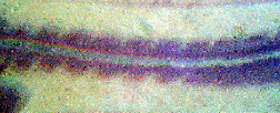
In the micrograph above taken longitudinally from a 72-hour chick
embryo, you can see the block-like somites developing from the
unsegmented mesoderm. The chick's head is to the left. The pale
line from left to right of the micrograph is the nerve cord.
- It is difficult to see where premyoblasts originate because
they look very similar to many other types of stem cells destined to
form other types of tissues.
Below is a diagram of a greatly simplified plan of a transverse section
through an
embryo. To use the same diagram for both farm mammals and poultry, the
bottom of the diagram only indicates the general
relationships of the somites
to the endoderm (gut), to the
coelom (body cavity) and to the parietal
lateral
plate mesoderm (ventral body wall). In other words, something
like the
top of the diagram may be seen in both mammalian and avian embryos,
whereas the bottom of the diagram is imaginary.
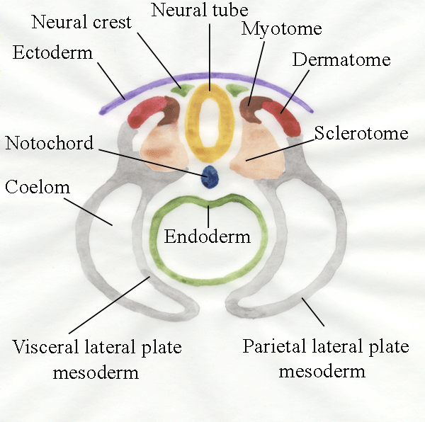
- Each somite is composed
of three zones around a cavity called the myocoele. The myocoele is of no
great importance and may simply be where the tissues come apart as they
are being prepared for microscopic examination.
- The sclerotome forms the
vertebrae.
- The myotome forms MUSCLE.
- The dermatome forms the
dermis (deep layer of the skin under the epidermis).
- The ectoderm forms the
epidermis of the skin.
- The endoderm forms the
gut and associated organs.
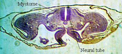 This
is what it really looks like!
This
is what it really looks like!
Genes. Seen in a whole embryo, each somite appears
as a cube of tissue, with left and right somites forming pairs in an
anterior
to posterior sequence. The division of continuous strips of
mesoderm to
become
somites follows a pattern created by the arrangement of cells early in
development,
Somite formation is determined by Pax
genes, while the
antero-posterior axis is determined by the Hox gene.
Mesenchyme cells. From
the Greek, this literally means cells 'poured' into the middle; in
other words, cells without any distinguishing features which fill a
space. More typically a histologist might say, "the sclerotome is
composed of loosely packed and morphologically undifferentiated
mesenchyme
cells." The mesenchyme surrounds the notochord and neural tube and
later
differentiates to form cartilaginous precursors of the vertebrae. In
farm
animals, myotomes become elongated from anterior to posterior, and this
obscures the original cuboidal shape of each myotome. Extensive changes
in the
shape and orientation of somitic cells occur during the development of
the
somites - in other words, the shape of the future animal is being
determined by the shapes of individual cells and they way they are
packed..
5.2 Origin of myoblasts
Premyoblasts
divide and their daughter cells become myoblasts (only some of them,
otherwise there would be no more dividing premyoblasts). Myoblasts fuse
to form myotubes. Myotubes
mature into muscle fibres. This
will be explained more fully in the
next lecture.
- The axial muscles of the body, including the tongue and
extraocular
muscles, are derived from the somitic mesoderm of the myotomes.
- Mesenchyme cells from both the somites and parietal layer
of the lateral plate mesoderm move into the limb buds, thus giving rise
to the muscles of the limbs (but there is plenty of debate about the
relative importance of somites and lateral plate in different animals)..
5.3 Limb buds
Limb bud formation is regulated by a
dialogue between ectoderm and mesoderm.
- The ectoderm makes
an apical ridge in response to a message from the mesoderm.
- The mesoderm
grows to form parts of the limb in response to a message from the
ectoderm.
- The mesoderm instructs the ectoderm to remain thick and to keep
inducing
mesodermal growth.
-
The mesoderm of the limb bud separates into
muscle‑forming and cartilage‑forming regions.
-
The chondrogenic
(cartilage-forming) regions can be detected
because they synthesize extracellular proteoglycan (the rubbery matrix
of cartilage).
-
Molecular differentiation is preceded by differential
vascularization (formation of blood vessels in different patterns).
Perhaps
vascular growth is involved in establishing metabolic gradients in
the developing limb?
-
Little
is known yet about the factors regulating the quantitative distribution
of
premuscle mesoderm and the potential for muscle growth in meat animals.
For
example, the maximum postnatal number of myofibres might be
predetermined
by the number of stem cells and by the number of times their
offspring
divide. At present it appears likely the numbers of mitotic
divisions in a
cell lineage are genetically programmed, but with environmental factors
controlling the rate of division.
-
The point at which multipotential stem cells (cells capable of forming anything) differentiate
to become myogenic stem cells (cells only able to form muscle) is
genetically regulated by a single gene, myd.
-
During the evolution of limbs in ancient amphibians, it
appears there was a primary division of the myogenic mesoderm into a
dorsal and a ventral mass. This still occurs in the embryos of our farm
animals.
Further information
W.H. Freeman & B. Bracegirdle. (1963). An Atlas of Embryology. Heinemann,
London. The original and still the best (in my opinion) manual to
identify what you are looking at in sections through the developing
chick.
B.M. Carlson. (1988). Patten's
Foundations of Embryology. McGraw-Hill Publishing, New York.
This is an updated version of Bradley M. Patten's "The Embryology
of the Pig" and the "Early Embryology of the Chick". These originals
are still easily obtainable at relatively low cost and have really nice
pictures and diagrams.
Structure and Development of Meat
Animals and Poultry, Chapter 6.